Wasp Wonders Under The Microscope: A Fascinating Look
Ever peered into a world so intricate, so detailed, that it redefines your perception of beauty? The microscopic realm of insects, particularly the often-feared wasp, offers a breathtaking perspective, revealing a complexity that rivals the most sophisticated engineering feats.
The common perception of a wasp might conjure images of a stinging menace, a pest to be swatted away. But under the lens of a microscope, these creatures transform into marvels of natural design. The compound eyes, the intricate mouthparts, the delicate wings all crafted with an elegance that belies their reputation. It's a journey into a world unseen, a testament to the power of observation and the endless wonders hidden in plain sight.
To fully appreciate this microscopic marvel, let's delve into the intricacies of studying these insects. Given their size, observing a live insect under a conventional microscope isn't ideal. A low-power stereo microscope, also known as a dissecting microscope, proves far more suitable. This type of microscope offers a three-dimensional view, allowing you to appreciate the surface details and structures of the wasp with greater clarity.
Consider the stinger, a weapon that sparks both fear and fascination. Viewed under a microscope, the stinger's intricate design comes into focus. The wasp's stinging apparatus is more than just a point; it's a collection of finely crafted lancets, often featuring tiny barbs that act as serrated edges. This detailed view reveals the remarkable engineering of a creature so often dismissed.
The following table provides a comprehensive overview of the tools and techniques used to study insects under the microscope, focusing on wasps as a prime example:
| Aspect | Details | Significance |
|---|---|---|
| Microscope Type | Low-power stereo microscope (dissecting microscope) | Provides a three-dimensional view, ideal for surface detail observation. |
| Magnification | Typically ranges from 10x to 100x | Allows for detailed examination of insect features, such as eyes, mouthparts, and stingers. |
| Specimen Preparation |
| Ensures optimal viewing conditions and preservation of the specimen. |
| Illumination Techniques |
| Enhances visibility of specific structures and features. |
| Key Features to Observe |
| Provides insight into the insect's anatomy, function, and behavior. |
| Mounting Medium | Euparal | Used for mounting insect specimens; maintains clarity and preservation. |
The wasp's head is a microcosm of sensory organs and formidable weaponry. Compound eyes dominate the view, each a collection of ommatidia, individual light-detecting units. The ocelli, three simple eyes often arranged in a triangle, provide additional visual input, aiding in navigation and orientation. The mouthparts, too, reveal their purpose designed for feeding and defense.
The study of wasps, and insects in general, under the microscope allows for comparative analysis. You can compare the stinger of a wasp to a needle, appreciating the natural engineering of the insect's defense mechanism. Observing the wings, with their intricate venation patterns and often iridescent scales, reveals the elegance of flight.
Consider a wasp building a nest on your balcony. Before removing the wasps, imagine taking one for closer inspection. With a stereo microscope, you could study the stinger, its design, and the way it functions. The process might involve dissecting the wasp, preparing a slide with mounting medium like euparal, and then viewing the specimen under varying lighting conditions: reflected, transmitted, and even ultraviolet light to reveal the fluorescence of certain structures.
The preparation of microscope slides offers another fascinating aspect. Making a dry-mounted slide is the simplest method. More complex techniques involve using a mounting medium and spacer rings. The resulting slides showcase the wasp's features in a way that a simple visual inspection never could.
The world of the microscopic wasp is not just about anatomy; it's about the beauty of design, the elegance of evolution, and the endless possibilities for scientific exploration. The iridescent abdomen of a cuckoo wasp (Hedychrum gerstaeckeri), for example, appears stunning under a microscope, showcasing the colors and textures often hidden from our vision.
The study also highlights the differences between wasps and bees. A wasp can extract its stinger and fly off, seemingly unharmed. In contrast, a bee loses its entire stinging apparatus, poison sac and all, when it stings a human.
The use of reflected, transmitted, and ultraviolet light can further enhance the viewing experience. Reflected light shows surface details, while transmitted light reveals internal structures. Ultraviolet light can cause certain structures, such as the eyes, to fluoresce, revealing even more details.
The images often shared in this field showcase the diverse applications of microscopic study, from studying the structure of the stinger to capturing the fluorescence of the compound eyes. The study of these insects is an exercise in observation and precision, a way to discover the beauty of nature.
The work of microscopists often makes the invisible visible, revealing the patterns, structures, and details that comprise these complex creatures. This also extends to the techniques for preparing specimens, such as making a dry mount slide and using mounting media.
The study can also extend to the practical side of insect identification and classification. The study might involve examining key morphological characteristics, such as the shape of the head, the arrangement of the legs, or the intricate patterns of the wings. This offers a way to explore biodiversity and the natural world around us, making it clear that there's a wealth of information available for those who are curious and willing to look a little closer.
The study of wasps under a microscope is more than a scientific endeavor; it's an invitation to observe the world around us with a newfound sense of wonder and appreciation. It is a reminder that even the smallest creatures are capable of remarkable feats of engineering and beauty. It is an invitation to explore and to be amazed, one tiny detail at a time.
For those eager to delve deeper, resources like Redbubble offer prints and apparel featuring these stunning microscopic images. Scientific communities, such as the Nikon Small World, showcase incredible microscopic images, highlighting the beauty and intricacies of the insect world. There are also individuals who document their microscopic explorations on platforms like TikTok, sharing their discoveries with the world.
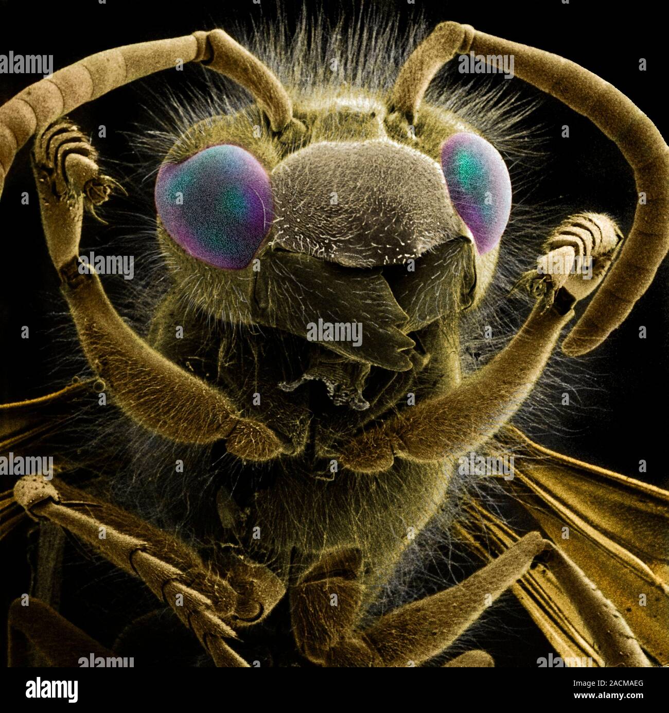
Common wasp (Vespula vulgaris), coloured scanning electron microscope
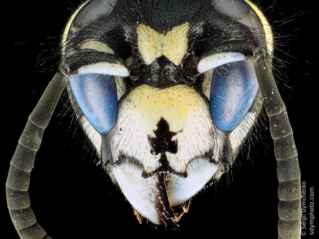
Wasp head under the microscope Sdym photo
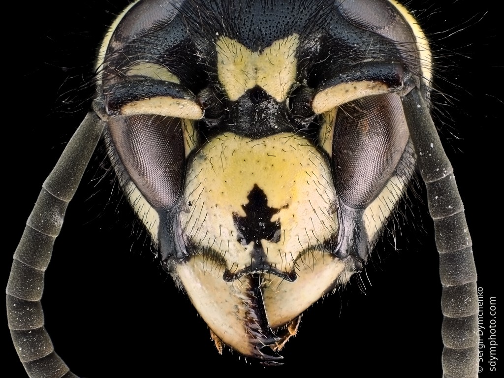
Wasp head under the microscope Sdym photo
